If you are searching about Necrosis observed within the tumor. | Download Scientific Diagram you've visit to the right web. We have 9 Images about Necrosis observed within the tumor. | Download Scientific Diagram like What Tumor Necrosis Factor (TNF) Has to Do With IBD, Gross morphologic and histopathologic changes. (A) Necrosis in tumor and also Tumor necrosis factor alpha (TNF) cytokine protein molecule, 3D. Read more:
Necrosis Observed Within The Tumor. | Download Scientific Diagram
 www.researchgate.net
 necrosis tumor observed cancer
Tumor Necrosis And Marked Cellular Atypical (HE, X40) Fig. 5
 www.researchgate.net
 necrosis cellular x40 atypical infiltration
Main Types Of Necrosis In Smooth Muscle Tumors. A, Tumor Cell Necrosis
necrosis tumor tumors leiomyosarcoma atypia cohesive infarct fascicles intersecting hematoxylin eosin hypercellular cytologic magnification
Necrosis Areas Within Tumor Shown By H&E Staining Observed With Light
 www.researchgate.net
 necrosis staining tumor observed
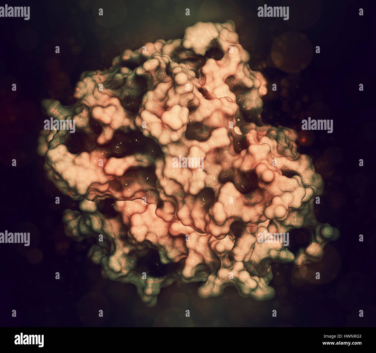
Tumor Necrosis Factor Alpha (TNF) Cytokine Protein Molecule, 3D
 www.alamy.com
 necrosis tumor factor alpha tnf cytokine molecule protein alamy 3d
Webpathology.com: A Collection Of Surgical Pathology Images
necrosis bone tumor cell giant webpathology infarct necrotic comments
Gross Morphologic And Histopathologic Changes. (A) Necrosis In Tumor
 www.researchgate.net
 necrosis tumor morphologic histopathologic necrotic ct26 eosin
Tumor Necrosis Factor Proteins Binding To Their Receptors On A Human
 www.dreamstime.com
 What Tumor Necrosis Factor (TNF) Has To Do With IBD
/15133-56a503d95f9b58b7d0da8fd5.jpg) www.verywellhealth.com
www.verywellhealth.com blood cells necrosis photomicrograph smear tumor factor cell granulocytes agranulocytes
Tumor necrosis and marked cellular atypical (he, x40) fig. 5. Tumor necrosis factor proteins binding to their receptors on a human. Blood cells necrosis photomicrograph smear tumor factor cell granulocytes agranulocytes
Tidak ada komentar:
Posting Komentar